Hi! This is my final project for ANT 4525 Human Osteology.
When considering what I wanted to do for my final project, I thought about what area of human osteology
I had the most trouble with.
By far, this was cranial anatomy. Because of the COVID-19 pandemic and quarantine,
I didn't have access to physical skull models. Reproductions of skulls were expensive, and while online 3D models
were helpful, I found that I wasn't fully grasping elements such as the orbits and how bones composed them.
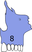
For this reason, I chose to make virtual jigsaw puzzles of the human skull from the anterior and lateral views. Hopefully, this will be helpful in understanding how bones appear from multiple angles.
On the right side of the screen, you'll find numbered definitions of each bone.
The numbers on the definitions correspond with the numbers on each bone for quick reference.
The puzzle pieces were edited in Clip Studio Paint and coded using CSS and HTML5. Free hosting is provided by Neocities.
To access the different puzzles, use the tabs at the top. If you would like to start over, refresh the page. :)
1.
Frontal Bone: Composed of the
squamous and
orbital parts. The
squamous portion encompasses the forehead. It is vertical and relatively flat. The
orbital portion includes the upper sections of the orbital and nasal cavaties. Notable features include the
supraorbital ridges (browridge) and the anterior portions of the temporal lines.
2.
Sphenoid Bone: Seated in front of the basilar part of the occipital bone. The
median portion contains the body of the sphenoid bone, which encompasses the
sella turcica and
sphenoidal sinuses. The
wings are comprised of two larger wings attached to the lateral sides of the body and two smaller wings on the anterior sides of the body. Articulates with the
frontal, ethmoid, temporal, parietal, vomer, and
occipital bones to form the middle-posterior section of the
orbits.
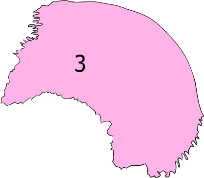
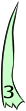
3.
Parietal Bone: Relatively large compared to other bones of the skull. Form the sides and superior areas of the cranium. Identifiable by the presence of
meningeal grooves on the inside surface, which branch out posteriorly towards the back of the skull.
4.
Ethmoid Bone: Divides
nasal cavity and brain. Found posteriorly between the
orbits, aligned with the
nasal bones. Articulates with the
frontal, sphenoid, nasal, maxillae, palatine, vomer, and
lacrimal bones.
5. Lacrimal Bone: Two small paired bones found on the medial wall of the orbit, near the front. Articulates with the frontal, ethmoid, maxillae, and inferior nasal conchae.
6.
Nasal Bone: Two paired bones which form the bridge of the nose. Articulate with the
frontal, ethmoid, and
maxillae bones.
7.
Zygomatic Bone: Paired bones which form the cheeks (cheekbones), as well as the lateral wall and floors of the orbits. Articulates with the frontal, sphenoid, temporal, and maxillary bones.
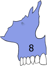
8.
Maxilla: Upper jawbone. Formed by two paired maxillary bones which fuse in the center. Contains the
maxillary sinus and anterior portion of the
hard palate.
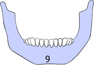
9.
Mandible: Bottom jawbone. Articulates with the
temporal bones.
10.
Temporal Bone: Form the
temples and contain components of the ears. Identified easily by
temporal line (which connects with the portion of the ridge seen on the
frontal bone) as well as a long, narrow
styloid process, though this often breaks off.
11.
Occipital Bone: Bone which forms the posterior base of skull. Identifiable by large hole known as the
foramen magnum (facillitates connection of medulla oblongata and spinal cord), as well as a raised cross-shape on the interior portion known as the
cruciform eminence.